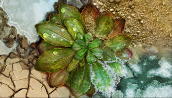
Mosaic image of a mouse brain slice improved by the software BaSiC. (Image: Tingying Peng / TUM/HMGU)
Research news - Today, tracking the development of individual cells and spotting the associated factors under the microscope is nothing unusual. However, impairments like shadows or changes in the background complicate the interpretation of data. Now, researchers at the Technical University of Munich (TUM) and the Helmholtz Zentrum München have developed a software that corrects images to make hitherto hidden development steps visible. When stem cells develop into specialized cells, this happens in multiple steps. But which regulatory proteins are active during the decisive branching on the development path? Using so-called time-lapse microscopy, researchers can observe individual cells at very high time resolutions and, using fluorescent labelling, they can recognize precisely which of these proteins appear when in the cell. Once a stem cell has been identified, it can be closely observed over several days using cell-tracking software. Yet, this 'surveillance work' often turns out to be difficult.
UM DIESEN ARTIKEL ZU LESEN, ERSTELLEN SIE IHR KONTO
Und verlängern Sie Ihre Lektüre, kostenlos und unverbindlich.