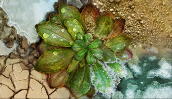
Detail of the brain vasculature of a mouse. The colors correspond to different diameters of the vessels. Red - largest diameters, blue - smallest diameters. Image: Paetzold / TUM, Ertürk/LMU Klinikum
Detail of the brain vasculature of a mouse. The colors correspond to different diameters of the vessels. Red - largest diameters, blue - smallest diameters. Image: Paetzold / TUM, Ertürk/LMU Klinikum - Coronavirus: University operations limited +++ make use of online services +++ many staff working from home - Biochemical methods and AI show even the finest capillaries - Diseases of the brain are often associated with typical vascular changes. Now, scientists at LMU University Hospital Munich, Helmholtz Zentrum München and at the Technical University of Munich (TUM) have come up with a technique for visualising the structures of all the brain's blood vessels - right down to the finest capillaries - including any pathological changes. So far, they have used the technique, which is based on a combination of biochemical methods and artificial intelligence, to capture the whole brain vasculature of a mouse. Changes in the blood vessels are a hallmark of numerous brain disorders - from traumatic brain injury to stroke.
UM DIESEN ARTIKEL ZU LESEN, ERSTELLEN SIE IHR KONTO
Und verlängern Sie Ihre Lektüre, kostenlos und unverbindlich.