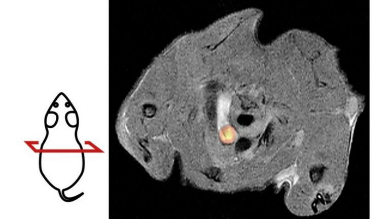
PET/MRI image of a mouse with atherosclerosis. The cross-sectional image shows the aortic arch as a bright structure in the center. The yellow glowing region represents an accumulation of potentially dangerous vascu-lar muscle cells. The drawing on the left illustrates the sectional plane of the PET/MRI image.
PET/MRI image of a mouse with atherosclerosis. The cross-sectional image shows the aortic arch as a bright structure in the center. The yellow glowing region represents an accumulation of potentially dangerous vascu-lar muscle cells. The drawing on the left illustrates the sectional plane of the PET/MRI image. Researchers at the University of Tübingen have developed a new method to better study atheroscle-rosis in mice. The non-invasive imaging method helps to better understand and treat narrowing of blood vessels, a cause of heart attacks and strokes. The new approach may also significantly reduce the number of animals used in experiments compared to previous methods.
TO READ THIS ARTICLE, CREATE YOUR ACCOUNT
And extend your reading, free of charge and with no commitment.