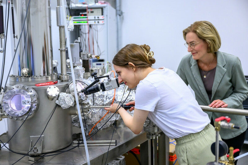
 PhD candidate Katarzyna Kwiecien (left) and Anika Schlenhoff at the scanning tunnelling microscope. They can use the device to visualise the individual atoms in the surface of a sample
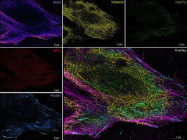
Five high-performance microscopes at the University of Münster. PhD candidate Katarzyna Kwiecien ( left ) and Anika Schlenhoff at the scanning tunnelling microscope. They can use the device to visualise the individual atoms in the surface of a sample © University of Münster - Michael C. Möller Many people are as fascinated by the microworld and the nanoworld as others are by the cosmos or the deep seas. These worlds seem to be inaccessible, apparently hidden from human eyes. However, across a wide range of disciplines, microscopes make it possible to take ever deeper and more precise looks at the smallest of details, and in ever higher resolutions - right down to atomic structures. The equipment which researchers use for this is highly complex and has almost nothing in common with the simple optical microscopes which many of us know from our schooldays. Fluorescence microscopy, for example, is a method which uses optical microscopy to label biomolecules within the specimen with fluorescent dyes and make the light they radiate visible.
UM DIESEN ARTIKEL ZU LESEN, ERSTELLEN SIE IHR KONTO
Und verlängern Sie Ihre Lektüre, kostenlos und unverbindlich.
Ihre Vorteile
- Zugang zu allen Inhalten
- Erhalten Sie Newsmails für Neuigkeiten und Jobs
- Anzeigen veröffentlichen